Case history:
A 54 year old male patient came with Pain in the left side of the chest radiating to the back side since 2 days, difficulty in breathing since two days.
Patient was apparently asymptomatic 2 days back then he developed pain in the left side of chest all over, of stabbing type, which increased on inspiration, radiating to the left upper back.
Pain ass with difficulty in breathing in breathing (a sense of difficulty/strain during inspiration).
No history of shortness of breath.
No history of palpitations orthopnoea, PND, headache, burning micturition, vomiting loose stools, cough fever
Patient has complete loss of vision in both eyes since 10years patient was apparently asymptomatic 12 years back then he developed severe dragging type of pain in both the eyes for which he went to the doctor & was diagnosed to have glaucoma & was given medication which he used for 2 years with no improvement.
He was also operated 10 years back due to loss of vision, but there was no improvement in vision despite surgical intervention, but the pain had improved.
There is no history of HTN DM epilepsy asthma CVA CAD.
GRBS values on presentation turned out to be 740mg/dL
No history of headache, tingling sensation, numbness.
No history of decreased urine output.
History of burning sensation of both feet since 1 year.
On Examination the patient was conscious coherent and cooperative.
Febrile -100F
PR 104BPM
BP 160/100mmHg
RS BAE+ decreased breath sounds in left ISA coarse crepitations in lt IAA
CVS S1 S2 heard no murmurs
P/A soft non tender
Investigations:
On day 1:
ECG at presentation:

ABG at presentation showed slightly decreased levels of pCO2

Random blood sugars elevated- 740mg/dL

Complete urine examination showed elevated sugar levels
Urine was also tested was ketone bodies which turned out to be negative.

Hemogram shows increased TLC- 19,800 & increased neutophil levels

Renal functions were mostly normal, phosphorus levels were slightly elevated in the patient

Serum osmolality was elevated- 324mOSM/kg

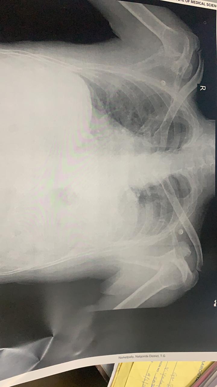
Chest X-ray on the first day shows slight effusion on the left lobe

Treatment:
9/6-
9pm- 538
10pm- 537
11- 472
10/6
12am- 437
1- 320
2- 266
3- 241
3:30- 229
4- 245
4:30- 274
5- 257
5:30- 317
6- 339 (1 unit of insulin in 39 parts of NS)
6:30- 435
7:30- 485
8:30- 446
10- 421
10:30- 413
11:30- 368
12:00- 431
12:30- 302
1:00- 346
1:30- 343
5pm- 316; 99.8F
7pm- 290
8pm- 1 unit subcutaneous insulin
10pm- 278
11/6 (GRBS Value in mg/dL)
12am- 238
2am- 179
4am- 164
6am- 157
8am- 271 (post breakfast)
9am- T.Glimiperide
10am- 264
12pm- 193; 101.2F
2pm- 203; 102.2F
4pm- 314
6pm- 319
8pm- 1/2 tab. Glimiperide
10pm- 361
12/6
1am- 268
4am- 254
6am- 238
8am- 238
9am- T.GLIMIPERIDE
10am-264
Fever chart:

Chest X-ray on day 4:

Culture sensitivity report showed klebsiella:

Chest radiograph and HRCT reports:


Pleural fluid- negative for malignant cytology;
Sediment smear was studied it showed scanty cellularity of Lymphocytes and few neutrophils only against eosinophilic proteinaceous background


Pleural sugar- 124mg/dL (elevated); pleural protein- 5g/dL, pleural LDH- 2240IU/L (elevated), pleural fluid ADA: 24U/L
Chest X-ray day 6:
USG- Chest shows thick septations and collapsed lung with minimal fluid
The ICD intervention would not be possible as there is less space
Consider breakage of loculations and septations through surgical intervention.


Bacterial culture negative for any aerobic bacteria.

Treatment:
1. Propped up position
2. Inj. piptaz 4.5gm/IV/TID day 7
3. Inj. Pan 40mg/IV/OD
4. T. Glimiperide BD (2.5mg - 1.5mg)
5. T. Ultracet 1/2 tab QID
6. BP PR RR hourly
7. GRBS 4th hourly
8. T vitC 1000mg/OD
9. T Telma 40mg/od
10. Inj neomol 1gm/iv infusion if temp >101F
11. Strict I/O charting
Diagnosis:
Left sided moderate loculated pleural effusion with left lower lobe pneumonic consolidation (exudative viral? Bacterial?)
Denovo HTN DM2
Cholelithiasis
Left sided moderate loculated pleural effusion with left lower lobe pneumonic consolidation (exudative viral? Bacterial?)
Denovo HTN DM2
Cholelithiasis
No comments:
Post a Comment